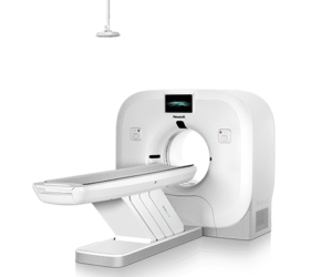
Description
Technical Specifications
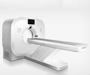
Magnet System
Technical Specifications
Magnet System
- 1.5 Tesla active shielded super conductive magnet.
- Should state the magnet length preferred Ultrashort 1.56 m
- It should have at least 60cm patient bore with flared opening; larger patient bore is preferable. The magnet should have facilities of better illumination, ventilation and designed to avoid patient claustrophobia
- The magnet should be shielded from the external interferences.
- The homogeneity of the magnet should be mentioned in relation to 10, 20, 30, 40, 50 cm DSV. Give details of the number of the planes plots and number of measurements per plane to measure the homogeneity. (A minimum of 50 cm DSV)
- Specify maximum FOV in all 3 axis (FOV to scan the largest possible human )40 cm minimum or more preferred)
- Specify Homogeneity at 50 (z) X 50 (x,y) cm DSV
- Specify the weight of the magnet including the gradient and covers etc.
- The front panel display at the magnet should display coil table position and also remote selection of coil elements,
Gradient System
- Actively shielded Gradient system with strength of at least 46 mT/m or more with the slew rate of 160 T/m/s or more. This slew rate of 160 T/m/s at 46 mT/m should be available in each axis independent, for overall better duty cycle performance of the gradient.
- The duty cycle should be 100 percent. Please give details.
- The Gradient system should have provision for eddy current compensation. (Bidder should demonstrate how they’ve compensated for the loss)
- Largest Field of View should be at least 50 cm in all three axis. Higher viable FOV will be preferred.
- Minimum TE in Gradient Echo 2D / 3D should be specified for all sequences. Minimum of 0.9 msec
- Minimum Slice Thickness in 2D should be specified. Minimum of 0.05 mm
- Minimum Slice Thickness in 3D should be specified. Minimum of 0.03 mm
- Maximum Echo Tr’ Length in both Spin Echo and Gradient Echo should be at least 256 or more.
- The measurement matrix should be from128x128 to 1024×1024 in both 2D and 3D imaging as well.
RF System
- RF system should be fully digital with transmit power of at least 18 Kw
- RF system should have at least minimum of 24 true independent RF receiving channels with each having independent ADC with bandwidth of 250KHz or more.
- Should have necessary hardware to support Phased array coils.
- Specify frequency stability and amplifier resolution.
- RF system should be compatible with parallel imaging techniques. It should be able to support time reductions with compatible coils in 2D/3D imaging in Body/ Neuro imaging up to factor of 2 or more
- RF amplifier should be solid state for overall better performance
RF Coils (How many coils are part of the equipment)
How many are to be purchased separately
N/B: All coils are needed/ essential for advanced detail of the MRI studies
- The main body coil integrated to the magnet must be Quadrature/ CP. In addition to this coil following coils should be quoted.
- Phased Array Head coil with mirror. It should be at least 24 Elements or more. Higher element coil will be preferred.
- In case above two coils do not suffice in combination for complete Neuro-vascular study from Aortic arch to Circle of Willis, please quote separate coil in addition to above two coils for this study. Please specify the max parallel imaging time reduction
- Phased Array Spine Coil for thoracic and Lumber spine imaging. Mention the number of coil elements available.
- It should be possible to do Head and Spine imaging together without changing the coil and the patient. It should be possible to do the same either with combination-of coils or a dedicated coil to achieve the same should be quoted
- Phased Array Body coil, capable of doing abdomen, pelvic, MRCP and peripheral imaging. It should have at least 12 elements and 45 cm FOV should be achievable.
- Flexible Coil -Large for imaging of large regions such as shoulder, hip and knee etc.
- Flexible Coil -small for imaging of small regions such as shoulders, wrist, elbow and ankle.
- Quadrature Extremity Coil for Knee Imaging
- Dedicated Shoulder Phased Array coil.
- Coil for Knee Imaging with 8 channels or more. Please specify the time reduction factor with parallel acquisition techniques.
- Bilateral Breast Coil, specify type and channel
- A coil for Neurovascular Application.
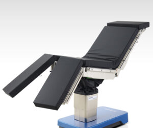
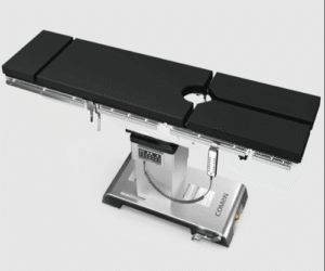
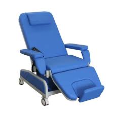
Patient Handling System: Dockable trolley/ stretcher system to limit lifting of patients into the examination couch. Also safe for patients
- The table should be fully motorized, computer-controlled table movements in up and down) (vertical) and horizontal directions. The position accuracy should be at least +/ – 1mm or better for higher reproducibility’ in advanced applications.
- The patient table should be able to withstand patient load of 200 kg
- The table should have facility for emergency manual traction in case of emergency. The table should have patient auto alarm system. .
- The CCTV/ Intercom system with LCD display to observe the patient.
- The table should deliver the protocols for automatic bolus chasing in peripheral angio with the automatic table movement.
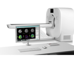
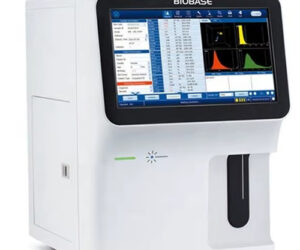
Host Computer /Main Console and Image Processor (Consideration for RIS and PACS to enable image transfer and archiving with enough storage capacity to archive images) consider cloud storage for unlimited space)
- Computer system should be latest in the industry, fast and efficient. It should have at least 16GB RAM.
- The main computer should have all the main processing software available in the Advanced workstation for quick review
- The system should have image storage capacity of at least 1 TB for at least 500,000 images in 256 x 256 matrix.
- The reconstruction speed should be at least 800 images per sec or more for full FOV 256 matrix and the image processor should have high RAM capacity of at least 16 GB for faster processing for advanced applications.
- The main Host computer should have at least 18-inch TFT type Color monitor. The main console should have integrated music system for the patient
- The system should have DVD and CD archiving facility (can add USB/ External drive) on the main console for storage of 50,000 images or more in 256 x 256 matrix. Additionally, 500 high storage DVD’s to be provided with external hard drives.
- One additional workstation with Color monitor to be provided for the applications as listed and 4 Additional workstations for concurrent interpretation (radiologists and academic
Application Software / Hardware (The range of purchased software will determine the capability of the machine e.g Diffusion, Perfusion, Spectroscopy, Tractography, BOLD, Cardiac, CSF flow studies, DWIBS
- The system should have basic sequences package with Spin Echo, Inversion Recovery, Turbo Spin Echo with high turbo factor of 256 or more, Gradient Echo with echo train length of 256 or more
- The application software for image smoothing and edge sharpness etc. for improvement in image resolution should be quoted and it should apply for major imaging applications.
- Single and Multi-shot EPI imaging techniques with ETl factor of 256 or more
- Fat and water excitation, please specify the application package
- Diffusion Weighted Imaging, with at least b value of 7000 (b-value used in diagnostics are 2000 or less. Higher b-values lead to loss in image resolution or more. The system (? Software) should have facility for ON Line automated calculation of ADC maps.
- Please specify the motion correction algorithm/package for high-resolution motion free diffusion weighed imaging with multishot/ segmented EPI techniques.
- It should be also possible to do 3D motion correction, please specify the application
8. BOLD imaging: BOLD. technique with automated 3D motion correction, z- score, and correlation analysis with color overlay on anatomical images. It should be possible to have Real Time Processing of BOLD imaging data on the main console for the complete reconstruction. ? software purchase - The perfusion and the BOLD imaging should be possible for the whole brain with motion correction techniques. Please specify the application package and the motion correction technique.
- Parallel Acquisition Techniques: Please specify the name of the package. It should have applications in abdomen, neuroimaging including diffusion and perfusion etc., free breathing abdomen imaging and Cardiac imaging. The scan reduction time should be mentioned.
- Bolus chasing with automatic moving table should be offered and should be available with fluoro triggered MR angiography for manual and fast switchover in less than 1 sec for ceMRA results.
- The system should be supplied with ECG Trigger; respiratory trigger, peripheral pulse trigger and external trigger Electrodes
- The system should have facility to do Head to Toe imaging without shifting the patient at one go for metastases study (DWIBS) and without any loss of SNR.
- The system should be available to perform Multi Direction Diffusion weighted imaging /Diffusion Tensor Imaging and the same should be possible on the main console.
- The system quoted should have image pasting software on the main console for ease of use.
- The system should be quoted with motion correction software for uncooperative Head patients, It should be possible to have the same routine in T1, T2 and FLAIR imaging.
- The system quoted should have the software for Whole Body Diffusion weighted imaging (DWIBS)
- The system should have acoustic reduction techniques to reduce acoustic noise to the lowest level. Please specify noise reduction technology and reduction amount.
- The system quoted should be able to do multi contrasts in a single image to save time.
Multiformat Dry Laser Imager
- Dry imager – DICOM 3.0 (or newer version) compatible, Dry chemistry. 600 DPI or more, with at least two film drawers. 14 x 17 “(35 x 45 cm) and 14 x14” (35 x 35 cm) size.
- System must provide complete batch filming with means to adjust image contrast and density.
- Imager must be controlled for exposure from the operator’s console and any workstation. An interlock/indicator system must be provided to prevent image production from one console, being intermixed with images from other consoles.
- Automatic transport system.
- Remote keypad, contrast inversion, 35mm adaptability.
- Should be connectable to multiple modalities like CT, MRI, Angiographic systems, Ultrasonography, with online PACS necessary interface provided. Filming must be possible with all modalities mixed on a film.
- Must be able to do serial processing imaging system wise when multiple systems are connected to the processor.
- All needed software and hardware must be provided
Accessories
- Patient Comfort Kit for different body parts (head, knee, shoulder etc) two for each to provide adequate support)
- MR compatible Injector (Dual head): Must have Independent dual Syringe power head and console must have full color touch screen with user-defined protocols with programmable interscan delay.
Environmental factors
- The unit shall be capable of operating continuously in ambient temperature of 300 C and relative humidity of 80%
- Chiller System
All the shielding requirements of the room will have to be done by the supplier.
UPS and Power supply
- Power input to be 220-240VAC, 50Hz, /440 V 3 Phase
- UPS of suitable rating shall be supplied for complete system with minimum 8 minutes backup
Voltage stabilizer with suitable rating will be supplied
Standards and safety
- Should be FDA and/or CE approved product
- Electrical safety conforms to standards for electrical safety IEC-60601 / ISO-13450
Warranty
1. 24 months from the date of satisfactory installation & handing over to the department.
Maintenance
Comprehensive maintenance contract (CMC) for the complete system will start after expiry of the warranty period. This will include replacement of spares including all consumables and sealed units, liquid Helium and labour. The contract will also include the recommissioning of the system in the event of magnet quench for whatsoever reasons. The maintenance contract will also cover comprehensive maintenance (Labour + spares) for UPS including batteries. Note that any Liquid Helium lost due to quenching or due to any other causes during the guarantee period shall be borne by the firm. System spare parts availability should be guaranteed for at least 10 years from the delivery of the system.
Documentation
- Two sets of operational/service/application manuals (Hard and soft copy) are to be provided along with the equipment.
- Detailed documentation on various sequences, spectroscopy, application software and evaluation software etc. are to be provided and the same must be updated regularly for next 10 years as and when these are released. (Timely software updates are key)
- Supplier is required to ensure mailing of product/research newsletters (on MRI and MRS) released from their R&D sites to the Hospital on a regular basis. This is to keep this centre abreast of the latest developments taking place in system technology and research techniques.
- The vendor is to provide a tender compliance sheet by giving all the necessary specifications, which should be supported by printed documentation sheets and certification of each item. In the absence of such documentation, a letter from the principals of the company should be provided.
Software up gradation
Software upgrades for the core system and all related applications for next 10 years to be provided free by the firm as a matter of routine as and when these are released, inclusive of minor hardware changes.